
Digital Radiology/ X-Rays
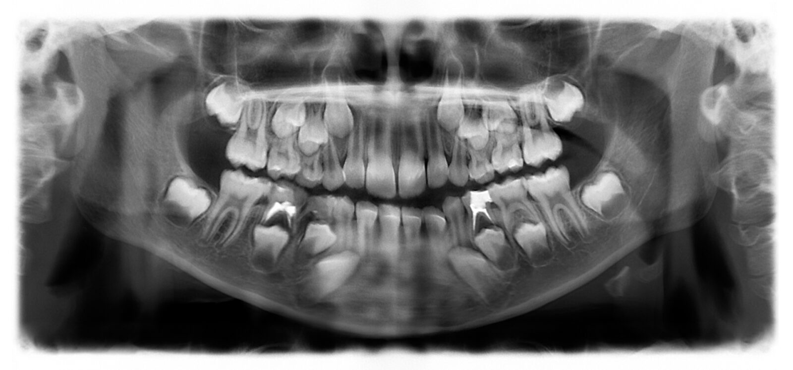
Dental x-rays are an important part of a complete dental exam. Annual x-rays for children in our office are 4-6 films. We may take a follow up film in 6 months depending on the patient’s need. X-rays are an essential diagnostic tool for finding hidden interproximal cavities
(cavities between the teeth), cysts, tumors, impacted wisdom teeth. They help guide determining eruption times of permanent teeth by the extent of root formation, and detect the presence of supernumerary teeth (extra teeth).
Children of all ages enjoy seeing their X-rays on the computer screen. It helps them to visualize their condition and gets them involved in
understanding the diagnosis or treatment. It is a great teaching tool. Modern dental x-ray machines are very safe. We use state-of-the-art, low radiation machines.
Intra Oral Camera
This wonderful new technology allows you to relax in your chair while simultaneously observing a real-time pictures of the inside of your mouth magnified beyond normal size on an adjacent computer monitor! Not only does this make it simple to see and understand what the doctor is telling you, but it makes it simple for us to keep incredibly accurate records from one visit to the next.
Diagnodent - laser assisted caries detection
DIAGNOdent aids in the detections of caries. Even very small lesions are detected at the earliest stage, enabling you to protect and preserve the tooth substance. The DIAGNOdent is significantly more accurate in identifying early stage lesions than conventional methods.
The KaVo DIAGNOdent has the unique ability to find “hidden caries”, a widespread phenomenon associated with the pervasive use of fluoride, which fortifies tooth enamel, However, despite the benefits of stronger enamel, traditional caries diagnostics, like the sharp explorer, once effective in the pre fluoride era, are no longer reliable. The implication of the current diagnostic challenge is that decay can progress under seemingly intact tooth structure, undiscovered and untreated by the clinician. The DIAGNOdent scans tooth structure with a harmless laser light, detecting sub surface caries. The device grades the lesion using a numeric scale, allowing the clinician to take appropriate action, before the decay can progress.
DIAGNOdent has received the ADA Seal of Acceptance which provides additional validation for the DIAGNOdent and demonstrates that the instrument has emerged as a new standard of care.
Sterilization and Patient Safety
Our office uses state of the art sterilization to ensure patient safety. Sterilization and disinfection are the basic steps in instrument processing and surface asepsis. Sterilization refers to the use of a physical or chemical procedure to destroy all forms of microorganisms, including the highly resistant spores.
We use Rapid Steam Autoclave at 275º F(35psi), for 15-20 minutes.
First, the instruments are prepared for the sterilization process. Patient debris and fluids are removed by placing the instruments in 3.2% glutaraldehyde for 40 minutes .Following this pre-disinfection step the instruments are transferred to an ultrasonic cleaner for another 15 minutes .Then the instruments are rinsed, dried, placed in self sealing sterilization pouches and sterilized in the autoclave. Instruments which can not be heat sterilized, are immersed in 2% glutaraldehyde for 10 hours to cold sterilize.
We use Biological, Chemical and Mechanical indicators to monitor our sterilization process.
Using bacterial spores to monitor the sterilization process is referred to as biologic monitoring (or spore-testing), and the bacterial spores used for monitoring the sterilization process are referred to as biologic indicators (BIs). Of the three methods, biologic monitoring is regarded as the most valid for monitoring the sterilization process, for it uses live, highly resistant bacterial spores. We biologically monitor our sterilizer once a week to ensure complete sterilization using spore strips and keep accurate records for our monitoring. These strips are enclosed in a glassine envelope and processed through the sterilizer. They are then sent to our spore testing center where they are tested for live spores.
Chemical monitoring involves using chemical indicators (CIs) that change color or form when exposed to specific high temperatures or to the sterilizing conditions within a sterilizer. This is referred to as chemical monitoring (or process monitoring). We use sterilization pouches that have special marking that change color when subjected to sterilizing temperatures.
Mechanical monitoring involves observing and recording the physical aspects (e.g., temperature, pressure, or time) of the cycle when the sterilizer is being operated. Our Sterilizer is serviced regularly to ensure proper functioning.
Barrier Controls – As recommended by OSHA and CDC our office staff wears protective eyewear, mask, and new gloves for each patient. For each patient light covers, head rest covers, suction tips, air water syringe tips , bibs and any item used that cannot be sterilized are discarded.
Disinfectants – These are used on chairs, counter tops, and other surface areas in all treatment rooms after each use.
Our goal is to provide the highest level of safety and comfort for our staff and patients. We are committed to staying current with the latest in infection control and sterilization guidelines. You can feel confident that your child’s health is protected in our office.
Dental radiographs or x-rays are an important part of a complete dental exam. A complete set of 18 x-rays for adults and 10 x-rays for children are usually taken at the initial dental exam. Thereafter 6-8 x-rays called checkup x-rays are taken every 6 -12 months depending on the individual needs. X-rays are essential diagnostic tools for finding hidden interproximal cavities, cysts, tumors, impacted wisdom teeth, determining eruption times of permanent teeth by the extent of root formation and presence of supernumerary teeth (extra teeth).
Digital Radiography, offers some positive advantages over typical film X-rays. The most remarkable is the reduction of radiation exposure, by as much as 80%, which makes dental X-ray taking safer and minimizes concerns about radiation exposure. You should know that 2 dental x-rays deliver 5,600 times less radiation to an unborn child than an upper GI series, 80 times less radiation than a chest x-ray, and 4 times less radiation than a normal day of background radiation playing in the sun. Dental x-rays are necessary and both safe and effective.
Digital imaging has been used by the medical community to make diagnostic information more accessible and more valuable. It is now available for dental offices. These dental x-rays are taken using electronic sensors that send the image directly to a computer. This image is displayed on a LCD monitor, can be enlarged, and can help the patient visualize and understand the doctor’s treatment recommendations more easily. It also faciliates the doctor’s diagnostic ability in “zooming in” and enlarging a specific area of the x-ray. The digital image only takes 10-15 seconds to appear on the monitor. A side benefit is that digital X-ray is also environmentally friendly. The sensors used do not contain lead foil, like conventional dental film, that needs to be recycled or disposed of as hazardous waste. Also, there are no chemicals or water involved in generating an image, reducing pollution and water consumption.
Digital X-ray generates pictures that are diagnostically equivalent to film based images. Many image enhancement tools allow many viewing options. They allow zooming, brightness and contrast control, reversing, colorization, and other features that can be used to assist the dentist in diagnosing dental problems. The images also can be transmitted electronically for either insurance purposes or to another doctor involved in treatment.
Kids really enjoy being able to actually see their X-rays on the computer screen. It helps them to understand their condition better and gets them involved in treatment. By placing a large image of an x-ray on a monitor that the child can see, we can begin to include the child in the diagnosis. It is a great teaching tool.
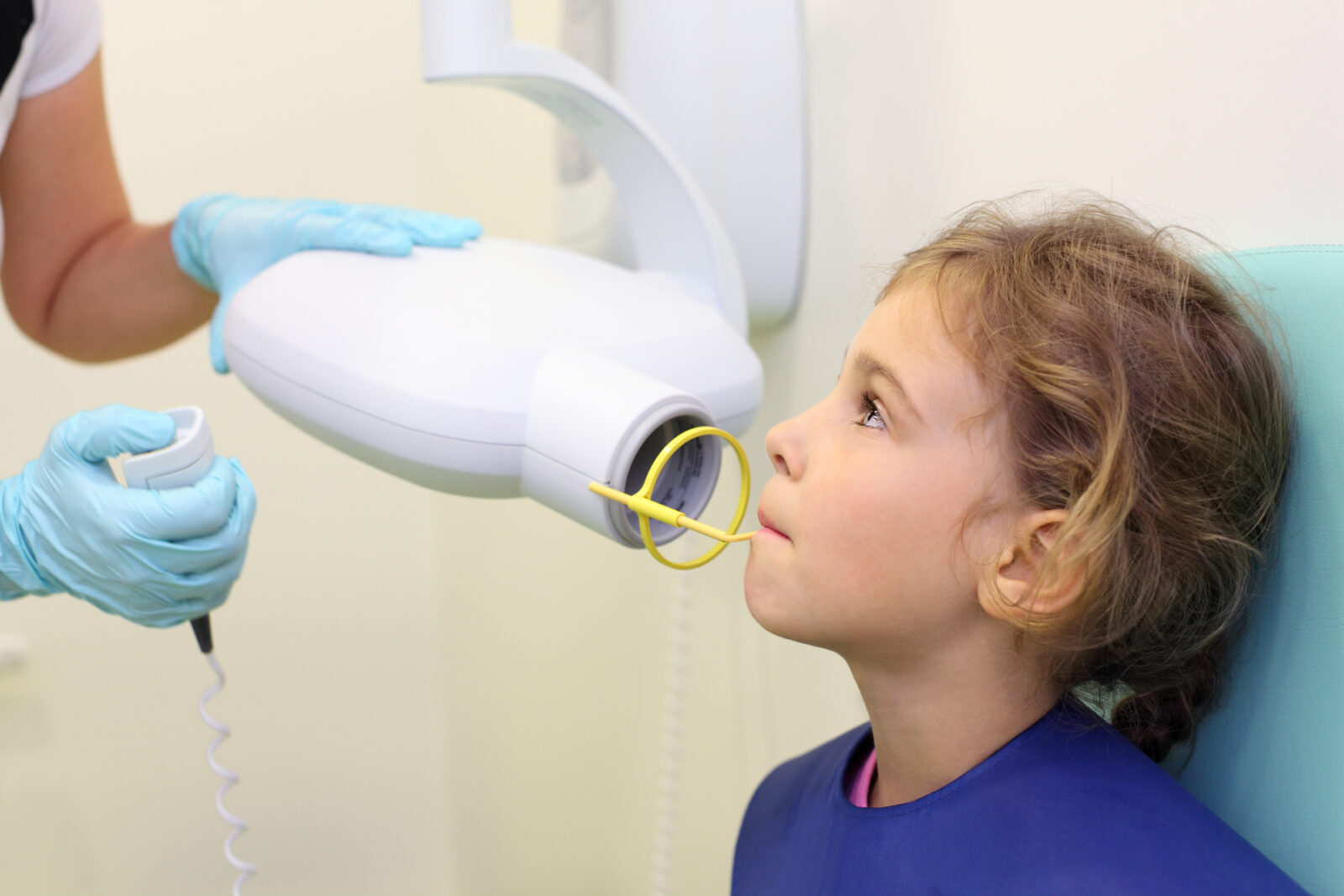
Taking the image:
A small, flat sensor is placed in the patient’s mouth next to the teeth. The sensor is connected to a computer by a thin wire. Next, an X-ray beam is sent through your teeth and into the sensor, which records the image of your teeth and sends it to the computer. The sensor can then be repositioned to capture other sections of the mouth.
This wonderful new technology allows you to relax in your chair while simultaneously observing a real-time pictures of the inside of your mouth magnified beyond normal size on an adjacent computer monitor! Not only does this make it simple to see and understand what the doctor is telling you, but it makes it simple for us to keep incredibly accurate records from one visit to the next.
DIAGNOdent aids in the detections of caries. Even very small lesions are detected at the earliest stage, enabling you to protect and preserve the tooth substance. The DIAGNOdent is significantly more accurate in identifying early stage lesions than conventional methods.
The KaVo DIAGNOdent has the unique ability to find “hidden caries”, a widespread phenomenon associated with the pervasive use of fluoride, which fortifies tooth enamel, However, despite the benefits of stronger enamel, traditional caries diagnostics, like the sharp explorer, once effective in the pre fluoride era, are no longer reliable. The implication of the current diagnostic challenge is that decay can progress under seemingly intact tooth structure, undiscovered and untreated by the clinician. The DIAGNOdent scans tooth structure with a harmless laser light, detecting sub surface caries. The device grades the lesion using a numeric scale, allowing the clinician to take appropriate action, before the decay can progress.
DIAGNOdent has received the ADA Seal of Acceptance which provides additional validation for the DIAGNOdent and demonstrates that the instrument has emerged as a new standard of care.
Our office uses state of the art sterilization to ensure patient safety. Sterilization and disinfection are the basic steps in instrument processing and surface asepsis. Sterilization refers to the use of a physical or chemical procedure to destroy all forms of microorganisms, including the highly resistant spores.
We use Rapid Steam Autoclave at 275º F(35psi), for 15-20 minutes.
First, the instruments are prepared for the sterilization process. Patient debris and fluids are removed by placing the instruments in 3.2% glutaraldehyde for 40 minutes .Following this pre-disinfection step the instruments are transferred to an ultrasonic cleaner for another 15 minutes .Then the instruments are rinsed, dried, placed in self sealing sterilization pouches and sterilized in the autoclave. Instruments which can not be heat sterilized, are immersed in 2% glutaraldehyde for 10 hours to cold sterilize.
We use Biological, Chemical and Mechanical indicators to monitor our sterilization process.
Using bacterial spores to monitor the sterilization process is referred to as biologic monitoring (or spore-testing), and the bacterial spores used for monitoring the sterilization process are referred to as biologic indicators (BIs). Of the three methods, biologic monitoring is regarded as the most valid for monitoring the sterilization process, for it uses live, highly resistant bacterial spores. We biologically monitor our sterilizer once a week to ensure complete sterilization using spore strips and keep accurate records for our monitoring. These strips are enclosed in a glassine envelope and processed through the sterilizer. They are then sent to our spore testing center where they are tested for live spores.
Chemical monitoring involves using chemical indicators (CIs) that change color or form when exposed to specific high temperatures or to the sterilizing conditions within a sterilizer. This is referred to as chemical monitoring (or process monitoring). We use sterilization pouches that have special marking that change color when subjected to sterilizing temperatures.
Mechanical monitoring involves observing and recording the physical aspects (e.g., temperature, pressure, or time) of the cycle when the sterilizer is being operated. Our Sterilizer is serviced regularly to ensure proper functioning.
Barrier Controls – As recommended by OSHA and CDC our office staff wears protective eyewear, mask, and new gloves for each patient. For each patient light covers, head rest covers, suction tips, air water syringe tips , bibs and any item used that cannot be sterilized are discarded.
Disinfectants – These are used on chairs, counter tops, and other surface areas in all treatment rooms after each use.
Our goal is to provide the highest level of safety and comfort for our staff and patients. We are committed to staying current with the latest in infection control and sterilization guidelines. You can feel confident that your child’s health is protected in our office.